


comprehensive Diabetes CARE


ON WHEELS
Bringing diabetes care to your doorsteps!


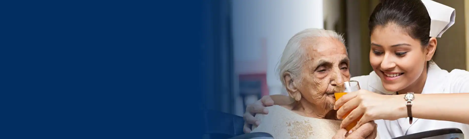
Healthy Ageing Clinic
Specialized in proactive, comprehensive care for diabetes and age-related health!

34 Years
10L+ Patients
34 Cities
51 Centres
Complete Diabetes Treatment under One Roof
We offer personalized, evidence-based care for individuals with diabetes, prioritizing their well-being.


Comprehensive support for adopting a healthy lifestyle to effectively manage diabetes and improve overall well-…

Diabetes can affect the eye in various ways. Diabetic Retinopathy is a common vision-threatening …

The foot is one of the major organs that can get affected by diabetes. The Foot Clinic at Dr. Mohan’s Diabetes …

In the headquarters of Dr. Mohan’s Diabetes Specialities Centre Hospital in Gopalapuram, Chennai, there is a 70…

At Dr Mohan’s, we keep pushing ourselves to extend the best care possible in the field of diabetes. By…

Dental gum problem (periodontal disease) is considered to be one of the major complications of uncontrolled…

The main aim of the pharmacy in the Dr. Mohan’s Diabetes Specialties Centre is delivery of authentic drugs to the …

Comprehensive support for adopting a healthy lifestyle to effectively manage diabetes and improve overall well-…


Eye Care at Dr. Mohan’s
Know MoreOur team of experienced ophthalmologists and optometrists are dedicated to providing top-notch care to ensure the health and well-being of your eyes. From routine power check-up, preliminary retinal examination to advanced surgical procedures, we are committed to offering the best possible treatments to our patients. At Dr. Mohan’s Diabetes Specialties Centre, your vision is our priority, and we strive to help you see the world more clearly. Trust us to take care of all your eye care needs with expertise and compassion.

Dr. V. Mohan
Chairman & Chief of Diabetology

My dream is to have a diabetes complication free world. At Dr. Mohan’s we strive to make people with diabetes live long & healthy life despite the disorder.
34
Cities Across India
10L+
Patients
8
States of India
130+
Doctors Team
35+
Services Offered
What Our Patients And Peers Think about Us

Excellent service.. All the facilities for the test purposes are available within the campus, which is very easy to complete all the test in a single go
A very good consultation by Dr Anjana mam and a very good follow up is being done


Sindhuja B
Chennai
One of the best places to go for your Diabetic review. They are very systematic, have all necessary facilities, the place is very clean and the staff are friendly. On the whole it is the best place one can go for Diabetic care.
Rajeev K R
Chennai
Staffs are very kind and their guidance are excellent. Taking extra care for each and clear the patient doubts in a polite manner. Especially environment is very neat and clean.
Ganesh Kumar
Chennai
Streamlined and smooth process, very professional and helpful stuff. There is always someone to guide you. You will definitely have good experience. Medical stuffs are polite and knowledgeable. No nonsense thing. You follow the process that they suggest, for sure you will come out of the hospital with a smile.
Been visiting here since last 10 years and still counting 😊
Manish Das
Chennai
Am happy with service of ms.mahalakshmi. excellent every time I meet her for review.
And other staff of other department is also kind and take care of my check up. here I have to specially convey my thanks to dr ramu who has taken of my health for past many years. once again I thank this centre for taking care of my health.
Siva Subramanium S
Chennai
Its an fabulous palace for treatment. The management is very comparative and well mannered and the staff also very helpful and kind towards the patient. I am grateful to came here for the treatment and what i want i get it from here. Thank you for your support and cooperation.
Biplab Debnath
Chennai
Dr Ramuu is always accessible to his patients, regardless of how far you live. His vast and in depth knowledge of diabetes have helped me manage my sugar levels better. The hospital itself has keen staff that makes communication easy.
Jane Kotian
Chennai
Very professional, very cheerful, very efficient. A happy place giving all good vibes. Have been bringing all my close relatives here for the last 20-25 years.
Siddhanth Ravindran
Chennai
Excellent customer care and help by the entire team at Dr. Mohan Diabetes Specialities centre.
My gratitude to Dr. Ranjit for his support throughout.
Mr. Krishnamurthy was very supportive and helpful for our requirements. Thanks
Balasubramaniyam KR
Chennai
Trusted diabetes care at
Dr. Mohan's Diabetes Specialities Centre
Book appointment with us today!